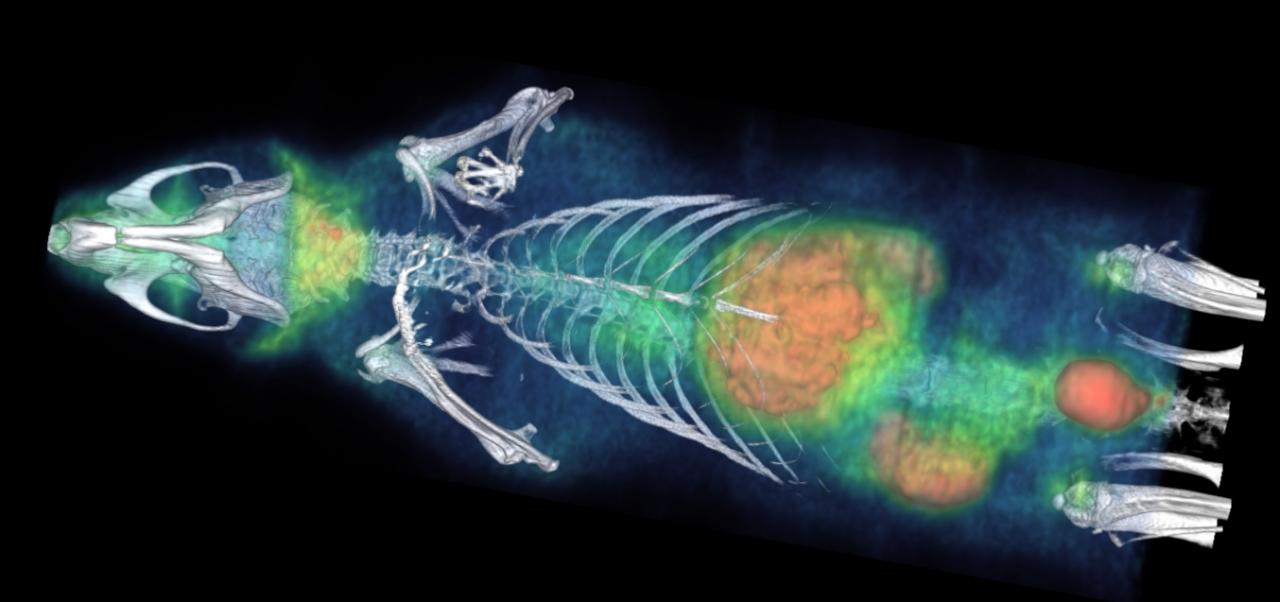
Preclinical molecular imaging
Imaging techniques are exceptional tools that allow researchers to acquire both, structural and functional in vivo information. Most importantly, these techniques are minimally invasive for the experimental animals, therefore ensuring the ethics of preclinical research.
The PET has special relevant characteristics:
It is a functional technique with high sensitivity (around nM) and it is minimally invasive nature. So, longitudinal (time course) studies in the same experimental animal can be carried out.
The high versatility of the PET allows the researchers to approach the study of multiple pharmacological targets.
The preclinical results from PET studies are of clinical translational nature and they can be further applied to the clinical context.
The detection process is based on the emission by the isotope of a positron that, when encountering a neighboring electron, undergoes a process called annihilation. This process generates two antiparallel high-energy gamma rays that are detected by the PET ring. By means of a subsequent reconstruction process, a three-dimensional tomographic image is obtained showing the distribution of the radiopharmaceutical.One of the main radiotracers used is the [18F]FDG because being a structural analogue of glucose allows for the evaluation of tissue metabolic activity. Furthermore, it is noteworthy that multiple pathologies and, especially those of neurological nature, are characterized by altered metabolic activity. Thus, the [18F]FDG has become a highly valued tool to characterize the metabolic profile of different types of neuropathologies that might further have predictive/diagnostic value as a marker as well as to evaluate their eventual pharmacological modulation.
In addition, a high-resolution CT module incorporated in the equipment complements the functional information within structural settings. Therefore, the PET-CT combinationprovides the information required for the anatomical localization of the functional alterations
Instrumentation
Staff
Rubén Fernández De la Rosa


